Ovēa Birth · Understanding Your OB Scan
Every pregnancy scan, explained
before you're on the table.
“Why did the room go quiet? Why can't the sonographer tell me anything?”
From the first heartbeat to the last check before birth, here's what each routine pregnancy ultrasound is for, when it usually happens, and what's actually being measured. Written by a high-risk OB sonographer who has performed thousands of them.
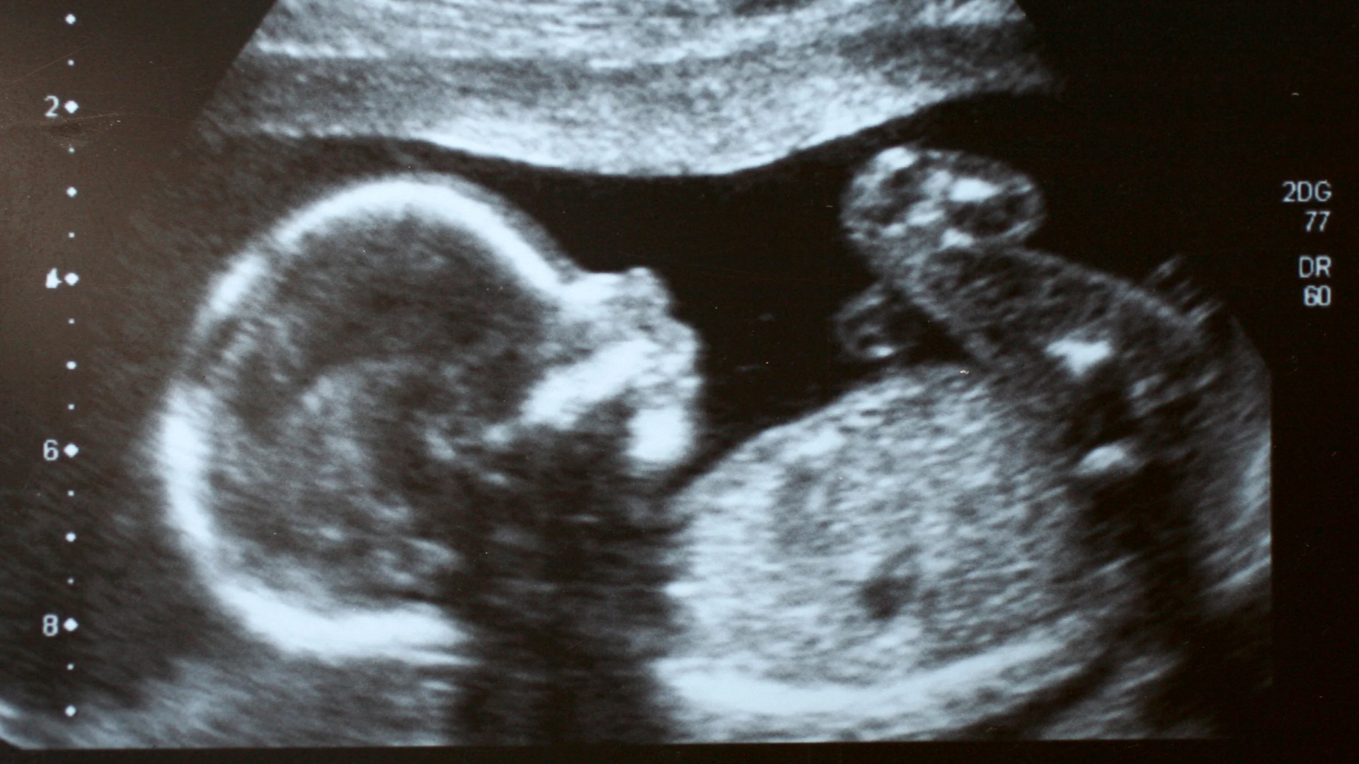
Your scan timeline
What to expect, and when.
Timing varies by provider, country and your own health history. Many pregnancies only need the first two or three. Growth scans and BPPs are added when there's a reason to look more closely.
“Is it in the right place? Is there a heartbeat? Is my due date right?”
Your first ultrasound answers the most important early questions. It's also the most accurate time to date a pregnancy: the crown-rump length (CRL, baby's length from head to bottom) in the first trimester is usually accurate to within about 5–7 days. If it differs enough from your last-period date, your provider may change your due date. That's normal and it matters, because every later measurement is compared against it.
What's being checked
- Location: the pregnancy is inside the uterus, not ectopic
- Cardiac activity: usually visible from around 6 weeks with a transvaginal scan
- Number of babies, and for twins, whether they share a placenta
- Size (CRL) to confirm or set your due date
- Your ovaries and uterus, such as cysts or fibroids worth noting
Transvaginal or abdominal?
- Transvaginal is used when the pregnancy is still very small (often before about 10 weeks), the view is unclear, the uterus tilts backward, or body habitus limits the abdominal view
- Abdominal works well once the uterus rises above the pubic bone. You may be asked for a fuller bladder.
- A transvaginal probe is safe in pregnancy and does not cause miscarriage. You can ask for a chaperone, or to insert it yourself.
“Do I need the NT scan if I'm already doing the blood test?”
The nuchal translucency (NT) scan measures a small fluid space at the back of baby's neck. Combined with your bloodwork and age, it estimates the chance of certain chromosomal conditions. Today, many clients skip it and choose more advanced NIPT (non-invasive prenatal testing) bloodwork instead. NIPT reads small fragments of baby's DNA that circulate in your blood, so it's a simple blood draw with no scan needed.
Common advanced NIPT options
- MaterniT 21 PLUS (Labcorp): screens for the most common chromosome conditions, including trisomy 21 (Down syndrome), 18 and 13, plus sex chromosome differences. It can also tell you baby's sex if you'd like to know.
- MaterniT GENOME: a broader screen that looks across every chromosome for larger missing or extra pieces of genetic material, not just the common trisomies.
- GENOME-Flex: if a MaterniT 21 PLUS result needs a closer look, the lab can re-run your saved sample with MaterniT GENOME's deeper analysis, usually without a second blood draw.
Some providers still recommend the NT scan alongside NIPT because it gives an early look at baby's anatomy, especially the heart, spine and abdominal wall, that bloodwork can't. Ask what your provider recommends for your situation.
“They kept going back to the heart. Should I be worried?”
This is the most detailed scan of pregnancy. The sonographer works through a checklist of structures, and some views take patience: the heart has several required angles, and the face and spine depend on how baby is lying. Being asked to walk, roll over or come back another day is very common. It usually means baby wasn't cooperating, not that something was seen.
Head & brain
Skull shape, the brain's ventricles, cerebellum and the fluid space behind it (cisterna magna)
Face
Profile, eye sockets, and the upper lip to check for a cleft
Spine
Viewed lengthwise and in cross-section, with the skin covering it
Heart
Four chambers, rhythm, and the major vessels leaving the heart (outflow tracts)
Chest
Lungs and diaphragm, with the heart sitting in the right position
Abdomen
Stomach, kidneys, bladder, and the abdominal wall where the cord inserts
Umbilical cord
Three vessels (two arteries, one vein) and where it attaches to the placenta
Limbs
Arms, legs, hands and feet: bones present and position
Placenta & fluid
Placenta location (low-lying or previa is rechecked around 32 weeks) and amniotic fluid amount
Cervix
Cervical length, sometimes measured transvaginally for the clearest view
Biometry
BPD, HC, AC and FL (head, belly and thigh-bone measurements) give an estimated weight and growth percentile
Sex
Only if you'd like to know. Just tell the sonographer before they start.
“They said baby is measuring small (or big). What happens now?”
Growth scans aren't routine for everyone. They're added when your provider wants to follow baby's size and fluid over time. The same core measurements as the anatomy scan (head, abdomen, femur) produce an estimated fetal weight (EFW) and a percentile. The trend across scans matters more than any single number. Ultrasound weight estimates can be off by around 10–15% in either direction, especially late in pregnancy.
Common reasons for growth scans
- Gestational or pre-existing diabetes: to watch for a larger-than-expected baby or extra fluid. Diabetes itself is diagnosed with a glucose test (usually at 24–28 weeks), not ultrasound.
- Suspected growth restriction (FGR/IUGR): estimated weight or belly measurement below the 10th percentile
- High blood pressure or preeclampsia
- Twins or more
- Fundal height measuring off, a previous small baby, or a low-lying placenta to recheck
What's measured
- BPD & HC: head width and circumference
- AC: abdominal circumference, the most sensitive marker of growth
- FL: femur (thigh bone) length
- Amniotic fluid: AFI or deepest single pocket
- Umbilical artery Doppler: blood flow through the cord, added when growth is restricted
- Position: head down or breech
A BPP checks that baby's environment is still a healthy place to be. It's common in higher-risk pregnancies, past your due date, or after reduced movement. The sonographer watches for specific behaviors for up to 30 minutes, often alongside a non-stress test (NST) of baby's heart rate. Each part scores 2 (seen) or 0 (not seen in the time).
| Component | What's being looked for | Points |
|---|---|---|
| Breathing movements | Practice breathing movements of the chest and diaphragm | 2 or 0 |
| Body movement | Several separate movements of the body or limbs | 2 or 0 |
| Tone | A limb or hand stretching out and flexing back | 2 or 0 |
| Amniotic fluid | An adequate pocket of fluid | 2 or 0 |
| Non-stress test | Heart rate rising with movement (monitor, not ultrasound) | 2 or 0 |
Bring these with you
Questions worth asking
at any scan.
Write them down beforehand. Appointments move fast, and it's easy to forget once you're on the table.
“Who will interpret my images, and when will I hear?”
Know whether results come from your OB, a radiologist or MFM, and how long it usually takes.
“Were you able to see everything you needed?”
If a view was incomplete, ask whether a follow-up is being booked.
“What percentile is baby measuring, and how does it compare to last time?”
For growth scans, the trend is the story.
“Is my placenta in a normal position?”
A low-lying placenta at 20 weeks often moves up. Ask if a recheck is planned.
“Can I have a copy of the report?”
Your report is yours. It's what an Ovēa Clarity Consult can walk you through.
Don't wait for your next scan if you notice…
- Baby moving less than usual, or a change in their pattern
- Vaginal bleeding, or fluid leaking
- Severe or constant abdominal pain
- Severe headache, vision changes, or sudden swelling of your face or hands
Call your provider or labor and delivery unit right away. For an emergency, call 911.
“You deserve to understand every image of your own pregnancy.”
— Ovēa · Women's Imaging Clarity
Have a report in hand?
Bring your scan.
I'll walk you through it.
A Clarity Consult is a personal 10–15 minute video walking you through what your pregnancy imaging shows, in plain language, with questions to bring to your provider. Or ask in the OB + Birth space inside Ovēa Circle.
Educational disclaimer: This guide is for educational purposes only and is not medical advice, diagnosis or treatment. Scan timing and practices vary by provider and region. Always follow the guidance of your OB, midwife or maternal-fetal medicine team. Hannah is an ARDMS-certified OB/Gyn sonographer and Certified Holistic Health Coach operating in an educational capacity. © 2026 Ovēa